The heart database: data
We provide data of the manual and automated segmentations.
You can download it following this link: HeartDatabase.tgz.
The file is 40Mb. md5sum is 8ff8a93358a77f1f29e34ae71b4ab281.
It is a tgz file, meaning you can extract it with tar -zxvf HeartDatabase.tgz.
Data format
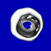
Each directory contains the data of a unique patient.Pat??/img/: contains the 3D image sequence, out???.pgm being the ???th 3D image of the sequence.
Pat??/expert1/ and Pat??/expert2/: contains the manual segmentation of the endocardial and epicardial border at end-systolic and end-diastolic time. An interior voxel is set to 255.
For each patient, a information file (Pat??/info.txt) contains the name of the image at end-systolic time (the manually-segmented one). The end-systolic time, manually-segmented image always corresponds to the first image of the sequence (i.e., out001.pgm).
The information file also contains the size of the original voxels (from DICOM), which is not the same from one patient to another. However, the provided data are isotropic, so this information is no longer pertinent.
Pat??/mse4d/: contains the automated segmentations for each time-step. As described in the paper, Pat??/mse4d/lvc/ contains the left ventricular chamber and Pat??/mse4d/lvcm/contains the left-ventricular chamber + myocardium.
Image format
The image format is an extension of the well-known PGM format, modified for supporting 3D.
Each image has a header of the form:
P7
100 100 14
255
On the first line, P7 states that this is a 3D PGM, each voxel taking one byte.
On the second line, 100 100 14 indicates that the image has 100 lines,
100 columns and 14 planes.
On the third line, 255 indicates that 255 is the maximum value of the
image voxels.
Following the header, the data is in 8-bit unsigned char raw format.
Site info
© 2006-2007 A2SI Lab.